
Área Pacientes
SI DESEA ENCONTRAR AL ESPECIALISTA MÁS CERCANO A SU DOMICILIO, PUEDE HACERLO EN LA PESTAÑA SUPERIOR «BUSCADOR DE PROFESIONALES».
♦ PATOLOGÍAS
♦ DICCIONARIO DE TÉRMINOS
♦ ASOCIACIÓN DE PACIENTES
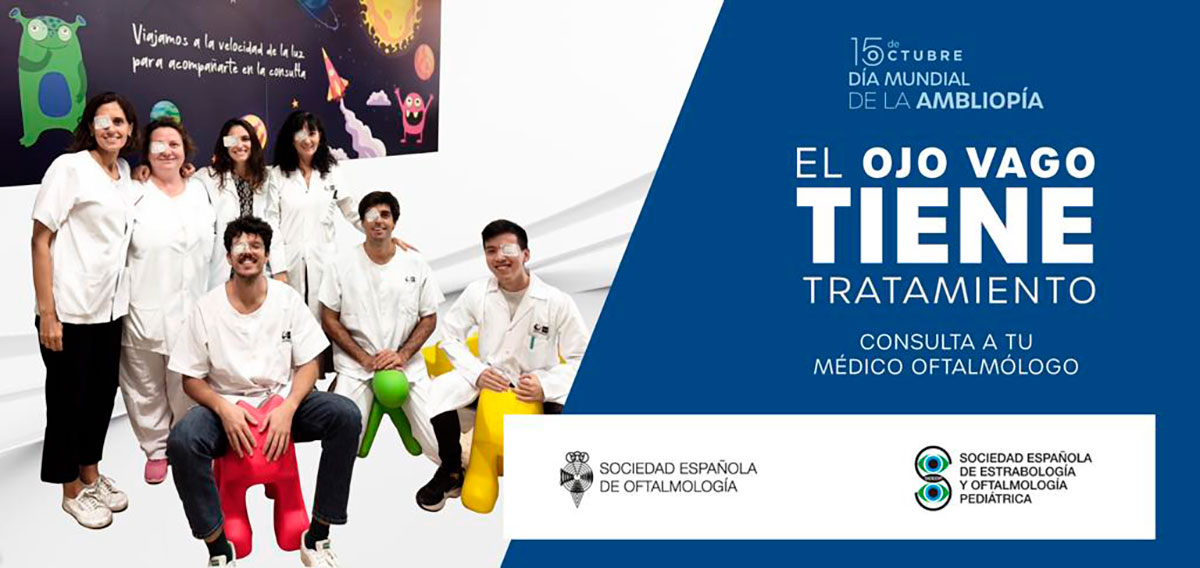
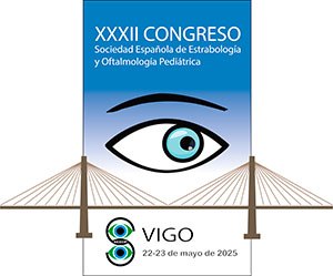
Eventos
PRÓXIMOS EVENTOS
Solicitud de certificados
 Boletín de solicitud de certificados de Congresos SEEOP
Área Científica
 Protocolos y Guías Clínicas
Subspecialty Exam in Strabismus and Pediatric Ophthalmology
Ya está abierta la solicitud del examen de la subespecialidad de Oftalmología Pediátrica y Estrabismo.
Todos los interesados podrán presentarse a través de este link
 Acta Estrabológica
A partir de este número, estará disponible para todas las personas interesadas en el mismo.
Acta Estrabológica n.2 2023 Julio-Diciembre
Acta Estrabológica n.1 2023 Enero-Junio
Acta Estrabológica n.2 2022 Julio-Diciembre
Acta Estrabológica n.1 2022 Enero-Junio
Acta Estrabológica n.2 2021 Julio-Diciembre
Acta Estrabológica n.1 2021 Enero-Junio
Acta Estrabológica n.2 2020 Julio-Diciembre
Acta Estrabológica n.1 2020 Enero-Junio
Acta Estrabológica n.2 2019 Julio-Diciembre
Acta Estrabológica n.1 2019 Enero-Junio
Acta Estrabológica n.2 2018 Julio-Diciembre
Acta Estrabológica n.1 2018 Enero-Junio
Acta Estrabológica n.2 2017 Julio-Diciembre
Acta Estrabológica n.1 2017 Enero-Junio
Si desean consultar los números anteriores deberán entrar en la sección de «Socios»
 Sociedades oftalmológicas y médicas
 Investigación
Encuesta IPOSC: Current Treatment Availability to Decrease Myopia Progression – IPOSC Global Survey
Registro de complicaciones de la inyección de toxina botulínica
Colabora con la investigación: Sd. Sturge-Weber
Registro nacional de Maculopatía por laser
Colabora con nosotros mandando tus casos a:
maculopatialaser@gmail.com
IPOSC -Myopia Survey. Action request
«Myopia is fast becoming a world epidemic requiring vigilant control of its progression.
We would like to compile data worldwide to better understand how we are currently meeting this condition. We would appreciate if you could spare a few minutes to reply to this survey»
Acceso al histórico de Acta Estrabológica exclusiva para socios

Contenido exclusivo para socios

Contenido exclusivo para socios
Contenido exclusivo para socios