CAPÍTULO 31 THE LYSOZYME AND THE LACTOFERRIN TESTS Otto-Paul van Bijsterveld |
| Flemming 1922 showed that tear, among
other biological secretions and fluids, has the remarkable property of dissolving certain
saprophytic cocci rapidly and completely. He obtained evidence that the agent concerned in
this lysis was an enzyme which he called "lysozyme". Even in high dilution he
found this enzyme to be able to lyse a certain coccus which he termed "Micrococcus
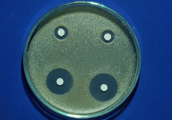
lysodeikticus". Certain other bacteria could also be lysed by lyzozyme. Lysozyme catalyses the depolymerisation of highly polymerized mucopolysaccharides. This means that it can be classified as a mucolytic enzyme, which breaks complicated mucoids into simpler forms, liberating in the process a reducing sugar believed to be hexosamine. The lytic effect of lysozyme on living and dead bacteria takes place by hydrolysis of mucoid substances found within the bacterial membrane. Lysozyme acts selectively on the cell wall of Micrococcus lysodeikticus by enzymatically cleaving the mucopeptide N-acetyl glucosamine (B 1-4) N acetyl muramic acid at the B 1-4 linkage. McEwen and Kimura used filter paper electrephoresis to study the lysozyme concentration in the tear fluid in normal persons and in patients with a variety of ocular diseases. In a unilateral case of dry eye as a result of trachoma no lysozyme was found in the tear fluid. Van Bijsterveld used a practical modification of the agar diffusion method using Micrococcus lysodeikticus as a substrate, to estimate the lysozyme contents of the tear fluid (figura 31-1). In his method he used filter paper discs of 6 mm diameter that were placed in the lower cul-de-sac to extract the tear fluid. He found the diameter of 21.5 mm of lysis to be the best limit between KCS and normal patients. The probability of misclassification was very low, i.e. 1% which suggested this test to be reliable in diagnosing dry eye as a result of tear gland degeneration.
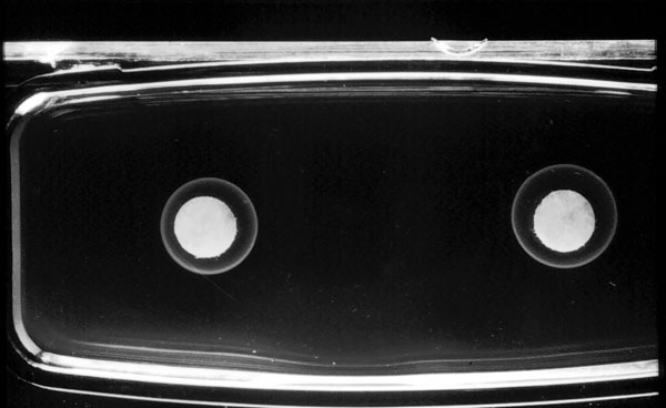
Lactoferrin, a transferrin like protein, previously reported to be present in bovine and human milk, was first shown to be present in the human tear fluid by Masson et al. There is some similarity between lactoferrin and transferrin. Their function is related to iron binding. The remarkable force of their metal binding possibly explains their bacteriostatic activity by making essential metal ions necessary for respiration and metabolism of micro organisms, unavailable Broekhuyse (1974) found that lactoferrin is a major tear protein and that transferrin is sometimes present in small quantities in the human tears probably because of leakage of serum protein in the tears. Gillette et al (1980) identified lactoferrin in most lacrimal tissues using the immunofluorescence technique. Lactoferrin was not found in the conjunctival tissues. They concluded that the primary source of lactoferrin in the human tears is the acinar epithelium of the main and accessory lacrimal glands. Radial immunodiffusion assay for lactoferrin is a more practical test than the radial immunodiffusion assay for lysozyme because of the high normal concentration of lysozyme in tears and the low antibody titer in antisera obtained commercially, and thus, a dilution step is essential for the lysozyme radial immunodiffusion test, while the lactoferrin radial diffusion test is a one step test. Also, the results of the radial immunodiffusion assay for lysozyme are somewhat influenced by an interaction between lysozyme and the agarose in the immunodiffusion plate. Janssen and van Bijsterveld (1983) introduced a simple, radial immunodiffusion assay for lactoferrin in tears (figure 31-2). This test does not require specific laboratory facilities and can be performed in the ophthalmologists office, and thus, provides an alternative for, or an addition to, lacrimal function tests currently used. The test is performed by collecting tear samples with filter-paper discs and placing them on previously prepared immunodiffusion plates.
|