ABSTRACT
In the course of the past 40 years, vitrectomy has gone from a high-risk pioneering procedure to a standardized surgery based on reliable equipment meeting high quality standards yielding foreseeable results. These developments were due to the creativity and perseverance of many expert surgeons as well as to the work of the bioengineering industry, frequently with the support of said surgeons. One of the latest developments in this field is the new Constellation® Vision System by Alcon, which includes the most recent breakthroughs in the development of vitrectomy gauges in different sizes: 20 G, 23 G and 25 G, fluidics, mechanized scissors and tweezers, xenon lighting, endolaser and phakoemulsification, in addition to a more comfortable user interface and enhanced management systems.
Key words: Phakofragmentation, fluidics, laser, xenon light, vitrectomy.
Pars plana vitrectomy (PPV) is a microsurgery procedure designed for eliminating vitreous gel, generally with the goal of accessing a diseased retina. The most frequent access is through threee separated incisions on the pars plana. Indications for surgery have increased together with the technical improvements. What may seem obvious at present is the result of a long struggle to achieve breakthroughs in vitreoretinal surgery through the experience and recommendations of a large number of posterior pole surgeons from all over the world as well as years of research by engineers to establish new standards of control and precision in dialy surgical practice.
INTRODUCTION
Going back in time, Machemer and Parel introduced in 1970 a rotating prototype marketed by Storz as VISC (vitreous infusion suction cutter) a couple of years later. This was a single port device that required large incisions and operated at low speeds. It was based on an electrical cutting system and aspiration through a syringe handled by an assistant. Subsequently, Ocutome appeared in the market, a lighter system developed by Connor O’Malley and Ralph Heinzun, which provided a pneumatic shearing system with a three-way operation. Ocutome provided aspiration control via a surgical pedal. It was followed by the Ocutome 8000 system developed by Carl Wang and Steve Charles. Its main contribution was the first linear suction, an integrated light source and a connected phako fragmenter.
A highly promising alternative which never reached the market was the OCM which even so set the groundwork for the Development of Accurus System® by Alcon Labs.
The main challenges for surgical material labs in the development of new vitreoretinal surgery systems is innovation and the addition of new functionalities. Alcon Laboratories’ Constellation® Vision System is the result of said approach. It includes an improved design of all OCM and Accurus System® technologies and also adds new capabilities which are described below.
CONSTELLATION® SYSTEM
The Constellation® Vision System was introduced in Spain at the Vitreo-Retina Congress on March 21, 2009. It probably will become the starting point for new systems yet to be released.
1. High performance Ultravit® vitrectomy probes
These are new vitrectomy pneumatic probes featuring ergonomic design and vertical cut. Three different gauges are available: 20 G, 23 G and 25 G with the option of utilizing higher shearing frequencies of up to 5,000 cpm in all gauges (fig. 1a y 1b). The Ultravit® probes are equipped with a new motor capable of actively advancing and reversing the shears. Two air lines provide microprocessor-controlled air pulses which allow the surgeon the freedom to select the working cycle of the probe best suited to the intervention to be performed (fig. 1c). The probe opening is arranged very close to the distal end, allowing work in the immediate vicinity of the retina to achieve enhanced precision vis-a-vis the vitreous (fig. 1d). In addition, the turbulences of the vitreous around the hand piece have been reduced to the minimum at any shearing frequency. Ultravit can be programmed in accordance with different vitrectomy modes, providing the traditional linear and 3D modes with an instantaneous mode.
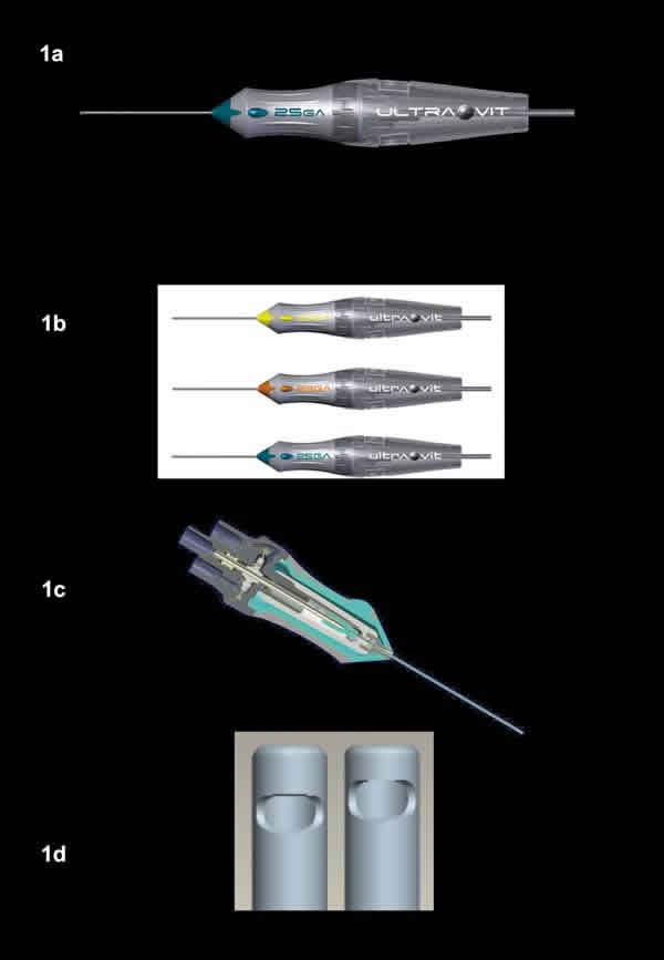
Fig. 1, a: Ultravit® probe. b: Availability of Ultravit® probes in 20 G, 23 G and
25 G gauges. C: The Ultravit® system includes two air lines for advancing and
reversing the shears instead of depending on spring system. d: The cutting
shears are arranged closer to the probe tip, allowing greater precision in the
vitreous operation.
2. Controllable duty cycle
The duty cycle is a time parameter that indicates the proportion between the opening and closing of the vitreotome nozzle. Until now the surgeon was only able to control the vacuum and the shearing frequency of the vitrectomy probe. The new Ultravit® technology, in which the nozzle opening and closing is carried out actively, allows a very precise control of the amount of aspired fluid independently of the desired shearing frequency, as well as of the preselected vacuum (fig. 2).
![]()
Fig.
2. Duty cycle comparison between Ultravit® in minimum and maximum
mode with Accurus®.
Three different duty cycles are available:
2.1. Central cycle: Maximizes the nozzle opening for central vitrectomy, where efficiency and higher flow rates are preferred.
2.2. 50/50 cycle: the nozzle remains open 50% of the duty cycle for surgeons who prefer equal opening and closing time.
2.3. Shaving cycle: Minimizes the nozzle opening for a soft tissue extraction where lower flow volumes and high safety are preferred.
Accordingly, the surgeon has a new tool for a precise tissue or membrane dissection from the retina surface or for cutting the vitreous or membranes in mobile retinas or under complex conditions.
3. Fluidics control and compensation
This is the main contribution of the system which allows the possibility of selecting a Venturi or peristaltic pum. The options are the following:
3.1. Vacuum control: Venture selected duty pump providing suction values of 50+ mmHg vis-a-vis existing Venturi equipment, reaching maximu values of 650 mmHg.
3.2. Vacuum control with flow limiter, which provides the possibility of selecting a fixed aspiration in cc/min independently of the density of the aspired fluids (extraction of silicone oil), avoiding possible hypotony effects. The Constellation® system regulates the amount to be aspired adapting the suction to the density of the aspired liquid (saline vs. silicone).
3.3. Flow control with vacuum limiter. When this function is activated, the main pump actas as a peristaltic pump separating the flow from the vacuum. This provides surgical standards adapted to the surgeons who utilize this pump in anterior pole procedures.
The Constellation® vision system comprises a number of ultrasound sensors capable of measuring fluid movements with the highest precision.
This allows for a balancing out of the pressure variations arising in the infusion line. In turn, this translates into a constant pressure in the ocular globe of ± 2 mmHg against the value shown on the equipment during surgery, thus avoiding the need to continuously monitor the infusion pressure as well as varying the height of the saline bottle during the intervention, leading to a reduction of ocular hypotony events (fig. 3).
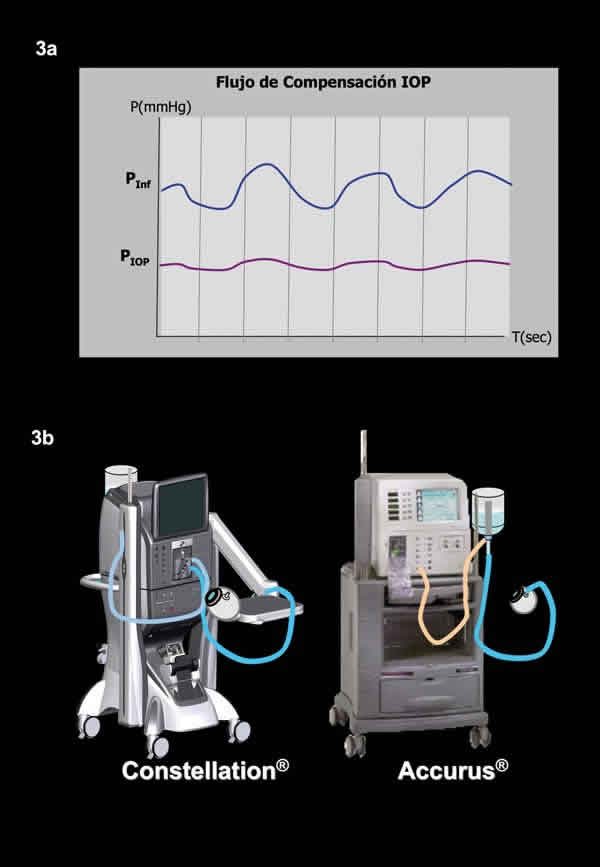
Fig.
3. a: Comparison of IOP oscillations. b: Comparison of Ultravit®
infusion system with pressurized cassette and in closed system with Accurus®
with non-pressurized cassette.
A new infusion flow limiter is included which provides an infusion flow reduction when we remove the active sclerectomy instruments (vitreotome, light fibre, tweezers, etc.) avoiding excessive fluid loss.
Other innovations, which enhance comfort and
safety, are:
3.4. VGFI system within cassette that allows
dropper exchanges without having to interrupt the surgery or having to prime the
system.
3.5. Immediate response to pressure changes.
3.6. Improved fluid stability during surgery.
3.7. Intraocular overpressure accessible from the pedal, with the option of measuring on the screen the exact overpressure time with alarm system and voice confirmation.
4. Advanced Xenon lighting
This system is used as a standard in all surgeries. The light
source is based on a long-
lasting xenon lamp (400 h) with a broad range of lighting probes and luminosity
indicated in percentages. This makes available bright light with optimized color
temperature to enhance visuali-
zation and less blinding. Now, working with lower gauge systems (23 G and 25 G) no longer limits the lighting power, as the system provides over 25 lumens with said gauges (fig. 4).
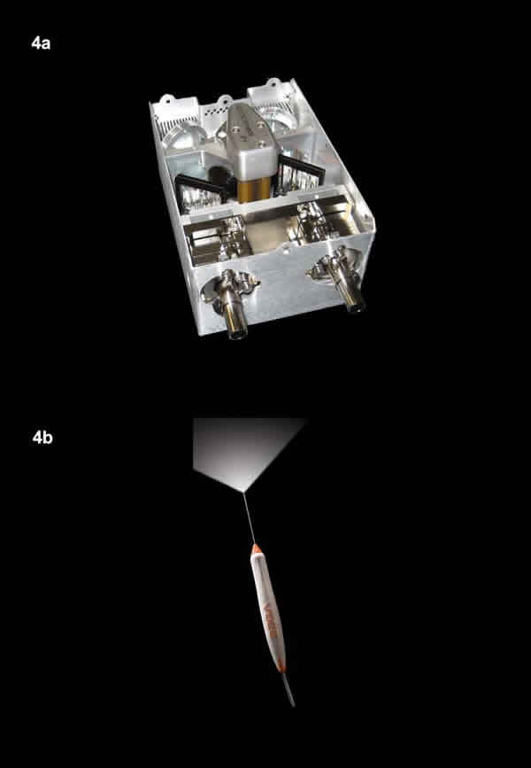
Fig.
4. a: The xenon lighting system provides good visibility while the dual lighting
allows the use of Chandelier® type lighting. b: The lighting angle
with the endo-lighting area exceeds 105º.
In addition, the system provides two light outputs with radiofrequency probe identifier (engauge RFID) which facilitates the use of the chandelier and torpedo-type field lighting system, with the option of an additional xenon light source.
5. Integrated Pure Point laser
The equipment comprises a 532 nm Purepoint green laser. All parameteres can be accessed from the surgery screen while a single surgical pedal controls the Standby/Ready functions as well as reducing and increasing the laser power.
In addition, safety is enhanced with the voice confirmation of the main functions. As with the lighting system, two laser output ports are provided and the laser parameters can be customized.
In order to simplify assembly and for avoiding mistakes, the probes are equipped with radiofrequency identification (RFID-EnGauge System®), and the range of gauges is of 20 G, 23 G and 25 G.
6. Additional functions
6.1. Fragmentation: a small, lightweight hand piece based on the Infiniti ultrasound torsional technology with a new priming system through suction or fluid filling, which allows a linear work mode specially designed for facilitating the emulsion of lenses or dislocated fragments to vitreous, with the pulsing fragmentation option.
6.2. Pneumatic scissors and tweezer system.
This is an air-drive handpiece which allows the use of disposable Grieshaber DSP
tips as scissors or tweezers. It operates in linear mode with the option of
pedal-controlled single or multiple cut.
In order to improve results and provide the advantages of manual systems, a pressure gauging functions is included to adjust the movement range of the scissors or tweezers.
6.3. Injection and extraction of silicone oil: the VFI mode provides air pressure for pedal-controlled injection and extraction of silicone oil through disposable 20 G and 23 G cannules.
A new possibility is offered by the combined mode which allows fast and simple fluid/fluid interchange.
6.4. Diathermia. By means of a high frequency diathermia (1.5 Mhz), the equipment provides a more superficial effect with fixed and linear diathermia modes. These modes are pedal-controlled as well and it allows for a very precise administration of the exact amount of diathermia to obtain the desired effect.
7. Innovative V-locity® functions
7.1. Automatic gas filling for endo-buffering with the option of two C3F8 and SF6 gas bottles. The syringe is automatically primed and filled with gas under sterile conditions in a few seconds in a concentration of 99.7%. From the main screen a conversion to cubic centimetres can be entered.
7.2. Automatic valve for fluid/air interchange, controllable from the screen or via the pedal, thus eliminating the three-step key.
7.3. Barcode scanner with reading of the disposable packs, which facilitates system assembly while updating stocks and providing input for billing.
7.4. Identification of radiofrequency connections (RFID-EnGauge System®) to eliminate the risk of erroneous connections by poorly qualified personnel.
7.5. Video help for assembly available in touch screen.
7.6. Articulated arm with tray.
7.7. Priming by fluid filling (push priming).
7.8. Active reflux from pedal with micro-reflux or linear mode of up to 120 mmHg.
7.9. Operated case report including all system metrics.
In summary, Constellation® Vision System
includes in a single console the latest breakthroughs and improvements of the
Accurus®, Infiniti® and laser Pure Point systems, besides
adding new developments in fluidics and vitrectomy probes, supplemented with
radiofrequency connections, videos and barcode reading to facilitate assembly
and stock control.