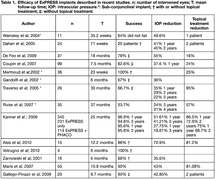
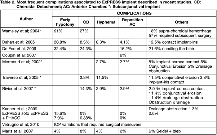
UPDATE REVIEW


Present and future of glaucoma ExPRESS implants

ARANA LARREA B1, PINAR-SUEIRO S2, CASTRESANA JÁUREGUI I1, MARTÍNEZ ALDAY N1

Ophthalmology Service, Cruces Hospital.
1 Graduate in Medicine. Ophthalmology Service. Cruces Hospital. Barakaldo. Vizcaya.
2 Ph.D. in Medicine and Surgery. Ophthalmology Service. Cruces Hospital. Cellular Biology Dept. Basque Country University (UPV/EHU).


INTRODUCTION

ExPRESS implants are a filtrating mechanism that maintains a permanent flow of aqueous humor in the subconjunctival and suprachoroidal spaces. These implants, made of stainless steel, have no valves and their penetration is limited by a distal flange and a proximal spur that prevents extrusion. The first ExPRESS device appeared in 1998, marketed by Optonol Ltd. The initial R-50 model was designed for insertion under the conjunctiva for chronic open angle glaucoma; subsequently, the P-50 model included an additional drainage perforation and a larger flange to prevent extrusion. At present, these implants are marketed by Alcon Labs. Initially, the implant was placed directly under the conjunctiva in a brief surgery of just a few minutes, although complications were detected together with frequent conjunctival erosions. Accordingly, Dahan and Carmichael (1) suggested a new approach, placing the implant under a scleral pad. This new technique is carried out in a similar way as traditional trabeculectomy, i.e., a subconjunctival incision is made on the base of the limbus and a pad comprising 50% of the overall scleral thickness. Before introducing the implant a pre-incision is made with a 27G needle and the ExPRESS device is introduced in parallel to the iris (figs. 1 and 2).


Fig. 1: ExPRESS implant correctly located in the anterior chamber with an active3 filtration bleb in a patient with limbar insufficienty.
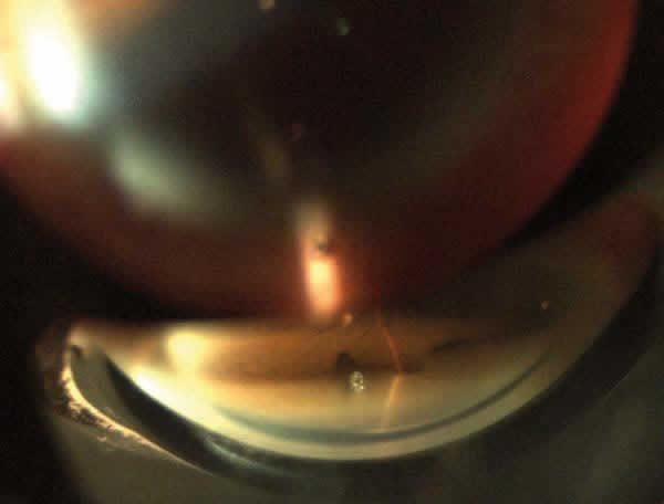

Fig. 2: Gonioscopy view of an ExPRESS implant placed in the anterior chamber parallel to the iris.

From a theoretical viewpoint, the ExPRESS implants have two main advantages: ease of insertion, which involves a significant reduction of surgery time, and a high degree of biocompatibility which prevents scar fibrosis, the most frequent cause of failure of filtrating techniques. Nyska et al (2) analyzed the histology of rabbit eyes submitted to ExPRESS implant surgery without finding signs of irritation or inflammation at 3 and 6 months, demonstrating their biocompatibility.

Since this new miniature implant was considered to be a reliable alternative to trabeculectomy, several studies have assessed a number of aspects such as efficacy, safety and side effects both in isolated filtrating surgeries and in combined surgery, or relative efficacy vis-a-vis other surgical techniques for glaucoma.

This literature review aims at analyzing the results described to date on the efficacy and post-surgery evolution of patients who underwent ExPRESS implants. To this end, we utilized the MEDLINE and EMBASE databases with the Pubmed and OVID SP search engines.


EFFICACY

In the literature we found efficacy studies on ExPRESS implants associated to other surgical techniques (phakoemulsification, vitrectomy or keratoplasty) as well as stand-alone surgeries (table 1).

As standalone surgeries, in a series of 11 eyes who had subconjunctival ExPRESS implants applied, Wamsley et al found 36% of failures. In the successful cases, a mean intra-ocular pressure (IOP) reduction of 49.6% was achieved. However, the complications rate turned out to be unacceptably high (3).

In what concerns the sub-scleral procedure, Dahan et al reported a series of 24 eyes with ExPRESS implants placed under a scleral pad. IOP reduction was of 41% at 1 year and 45% at 2 years (1). In a series of 37 eyes, De Feo et al (4) obtained IOP reductions of 55% after a mean follow-up of 18 months, with a success rate of 78% (IOP < 18 mmHg without topical medication) and a success rate of 70% with IOP < 15 without topical medication. Overall, 16% of patients required topical treatment to control glaucoma after receiving the implant. Finally, Coupin et al (5) analyzed 99 eyes with larger implants under a scleral pad and described IOP reductions of 37.6% after one year follow-up. The absolute success rate amounted to 62.6% and 24% of operated patients continued with topical treatment.

Various studies analyzed the efficacy of the combined technique comprising phakoemulsification with the ExPRESS implant under the conjunctiva. Accordingly, Mer-moud (Mermoud A, Rivier, E. The ExPRESS miniature glaucoma implant in combined surgery with cataract extraction: a prospective study. Paper presented at: Annual Meeting of the Ame-rican Academy of Ophthalmology; October 2002, Orlando, Florida, USA), in a retrospective study on 36 eyes, demonstrated that of the 6 patients who reached 12 months follow-up, 100% maintained IOP values ≤ 21; 2 of the 6 patients required topical anti-glaucomatous treatment during follow-up. Gandolfi et al. (6) achieved an IOP reduction of 36% with the probability of maintaining IOP < 16 mmHg after 9 months of 67% . In a series of 26 eyes, Traverso et al. (7) reported an IOP reduction of 35% at one year, 29% at 2 years and 22% at 3 years, with a success rate of 66.7% at 2 years. Reduced use of drops was of 95% at one year. Rivier et al (8) analyzed 35 eyes in order to reduce topical medication required to control glaucoma; they achieved 57% success in their initial objective, with 32% of patients maintaining IOP < 18 mmHg without topical hypotensor treatment. They also observed that the number of patients requiring topical medication and the dosage thereof increased with the passage of time and they pointed out that topical treatment it sends+++ is one of the causes of implant failure due to the irritation they produce on the ocular surface. Even so, they obtained an IOP reduction of 24% at 3 years and 31% at 4 years albeit with a relatively high complications rate.

Kanner et al (9) reported the most complete retrospective study, comparing the application of isolated ExPRESS implants with implants associated to cataract surgery with phacoemulsification and after the application of mitomycin C (0.4 mg/ml) during 2 minutes. The study comprised 345 eyes; 231 with ExPRESS implant on its own and 114 eyes combined with cataract surgery. The mean follow-up time was of 25 months. In the ExPRESS implants group on its own, the surgical success rate was of 96.9% at one year and 94.8% at 3 years. They defined this success as maintaining IOP between 5 and 21 mmHg with or without topical ocular medication. In this first group, the IOP reduction percentage was of 51.61% after one year follow-up and of 41.21% when follow-up was completed after 3 years. In the combined surgery group, the success rate was of 95.6% after one year, maintaining the same rate after 3 years. The IOP reduction was of 27.75% at one year and 19.61% at 3 years. The study concluded that IOP reduction was higher in the standalone ExPRESS implant group than in the combined surgery group. However, it must be taken into account that the baseline IOP of the first group was higher. The reduction in the number of drops required was statistically significant in both groups, but the number of pre-surgery drops required in the second group was significantly lower and the number of drops needed after surgery was significantly higher in the second group after one year of follow-up. Accordingly, the reduction of medical treatment was higher in the standalone ExPRESS implants group. No differences were observed in the postop complications between both groups.

In what concerns the ExPRESS implant combined with other techniques, we found in the literature the study analyzing a series of 15 eyes that required the application of on ExPRESS implant after penetrating keratoplasty, achieving a success rate of 86%. The number of drops required diminished significantly and edematous corneal grafts improved with IOP reductions (10).

Michele Vetrugno et al (11) implanted the ExPRESS device under the sclera in for previously vitrectomized cases. The complications they encountered were IOP variations that required simple surgical maneuvers. All cases had satisfactory control at month 6.

There is a variety of studies comparing this emerging surgical technique for managing glaucoma with more extended techniques such as non-perforating deep sclerectomy (NPDS) and trabeculectomy.

Zarnowski et al. (12) compared said technique with NPDS and found a number of infrequent complications in both groups as well as comparable efficacy rates at 6 months.

In 2006, Maris et al (13) compared 50 eyes submitted to trabeculectomy after ExPRESS implant under scleral pad, dividing also the group between single surgery and combined with cataract surgery. Even though results suggested better tension control in the group of patients submitted to trabeculectomy after 3 months follow-up, IOP became equal in both groups after 3 months . The mean IOP reduction was of 40-46% in the group of ExPRESS implants and of 28-45% for the group of trabeculectomy patients.

In what concerns the number of post-surgery medication required, the reduction was similar. In contrast, a higher number of complications were observed in trabeculectomy patients, such as early hypotony and choroidal detachment. It must be emphasized that in this study the

baseline IOP was lower in the patients who underwent combined surgery involving phacoemulsification and lens implant as though the final results as regards IOP control was similar in both groups.

In 2008, Gallego-Pinazo et al (14) carried out a similar study with 20 eyes in each group and obtained a success rate of 90% with ExPRESS against a 2% with trabeculectomy. The analysis did not demonstrate differences in long-term post-surgery IOP, as though in the first 2 weeks they observed that IOP figures were lower in the trabeculectomy patient group.


COMPLICATIONS

it is important to differentiate whether the implant is inserted under the conjunctiva or the sclera, because the majority of complications are described in sub-conjunctival implants.

The most frequent complications described were conjunctival erosion, early hypotony, hyphema, choroidal detachment, separate choroidal hemorrhage, implant rotation or filtration obstruction (table 2).

In what concerns subconjunctival implants, Wamsley et al. (3) described in a series of 11 cases 36% of failures, 91% of early hypotony, 27% of choroidal detachment, 18% of supper choroidal hemorrhage and 57% of cases that required subsequent re-interventions. However, other authors describe the presence of these complications with lower frequency rates. For instance, Traverso et al (7) presented 23% of early hypotony, 11% of hyphema, 7% of implant rotation and 11% of conjunctival erosion (fig. 3) in 26 eyes, while Mermoud et al referred 28% of cases requiring re-interventions, 3% of hyphema, 3% of hypothalamia and 8% of synechiae or contact with the cornea. In turn, Rivier et al. (8) reported 28% of major complications that required implant removal and an additional 28% of minor complications that did not require re-interventions. In this series, the follow-up time was higher, comprising 3 years.


Fig. 3 (A and B): ExPRESS implant inserted at the sub conjunctival level in a patient who underwent penetrating keratoplasty due to keratocone (A). showing conjunctival laceration of the overlying conjunctiva associate to incipient calcium deposit at that level (B), that required immediate withdrawal.

In the group of ExPRESS implants placed under a scleral pad, complications are described with less frequency. Accordingly, Dahan et al (1) and De Feo et al (4) described 20% and 32% respectively of hypotony and 8% and 24% of choroidal detachment. Coupin et al (5) found only 6% of athalamia cases, without finding any case of infection, extrusion or erosion in a series comprising 99 cases and of follow-up period of 7.5 months. Kanner et al (9) compared a series of 345 eyes with 4% of drainage obstruction resolved with YAG laser, 23% of early hypotony and only one case of hypothalamia and only 2 cases of implant exposure or dislocation after a mean follow-up period of 26 months.

The literature also includes reports of isolated cases of implant dislocation into the anterior chamber in the third week (Teng et al., 2008) (15) and of spontaneous extrusion after 2 years (Tavolato et al., 2006) (16). After a follow-up period of 10 months, Maris et al (13) published a table comparing the complications in the ExPRESS group under a scleral pad and trabeculectomy group that respectively referred for percent and 32% of early hypotony, 8% and 38% of choroidal detachment, 4% and 4% of hyphema and 2% and 0% of endophthalmitis. In a similar study, Galle-go-Pinazo et al (14) reported 30% of hypothalamia, 40% of hyphema and 15% of choroidal detachment in the trabeculectomy group and did not find cases with these complications in the ExPRESS implant group.

The last 2 studies referred above demonstrate a considerable reduction of post-surgery complications in comparison with traditional trabeculectomy.

Of all the analyzed studies, only one had a mean follow-up time of 3 years. Of the remainder, two reached 2 years follow-up and the rest had shorter periods, which evidences of the need of studies involving longer follow-up periods.


CONCLUSIONS

The studies conclude that the ExPRESS implant is an efficient technique in what concerns tension control, comparable to other surgical techniques for glaucoma. Its insertion under the conjunctival its controversial because the number of complications increases. However, when the device is inserted under a scleral pad, the risk of complications diminishes significantly, to the point that reports describe a lower complications risk in the immediate post-surgery period than in patients intervened with trabeculectomy.

The combination of implant with the phacoemulsification, penetrating keratoplasty or vitrectomy also appears to be safe and efficient. Specifically, the data discussed in this paper allow us to deduce that patients with a higher risk of sudden hypotony after of filtrating procedures such as trabeculectomy (i.e., aphakic or vitrectomized patients) could benefit from this technique.


REFERENCES


  1. Dahan E, Carmichael TR. Implantation of a miniature glaucoma device under a scleral flap. J Glaucoma 2005; 14: 98-102.
  2. Nyska A, Glovinsky Y, Belkin M, Epstein Y. Biocompability of the Ex-PRESS miniature glaucoma drainage implant. J Glaucoma 2003; 12; 275-80.
  3. Wamsley S, Moster MR, Rai S, Alvin HS, Fontanarosa J. Results of the use of the Ex-PRESS miniature glaucoma implant in technically challenging, advanced glaucoma cases: a clinical pilot study. Am J Ophthalmol 2004; 138: 1049-51.
  4. De Feo F, Bagnis A, Bricola G, Scotto R, Traverso CE. Efficacy and safety of a steel drainage device implanted under a scleral flap. Can J Ophthalmol 2009; 44: 457-62.
  5. Coupin A, Li Q, Riss I. Implant miniature Ex-PRESSTM placé sous un volet scléral Dans la chirurgie du glaucome á angle ouvert: étude rétrospective. J Fr Ophthalmol 2007; 30: 1:18-23.
  6. Gandolfi S, Traverso CF, Bron A, Sellem E, Kaplan-Messas A, Belkin M. Short-term results of a miniature training implant for glaucoma in combined surgery with phacoemulsification. Acta Ophthalmol Scand 2002; 236: 66.
  7. Traverso CE, De Feo F, Messas-Kaplan A, Denis P, Levartovsky S, Sellem E, Badalà F, Zagorski Z, Bron A, Belkin M. Long term effect on IOP of a stainless steel glaucoma drainage implant (Ex-PRESS) in combined surgery with phacoemulsification. Br J Ophthalmol 2005; 89(4): 425-9.
  8. Rivier D, Roy S, Mermoud A. Ex-PRESS R-50 miniature glaucoma implant insertion under the conjunctiva combined with cataract extraction. J Cataract Refract Surg 2007; 33: 1946-52.
  9. Kanner EM, Netland PA, Sarkisian SR, Du H. Ex-PRESS miniature glaucoma device implanted under a scleral flap alone or combined with phacoemulsification cataract surgery. J Glaucoma 2009; 18: 488-91.
  10. Ates H, Palamar M, Yagci A, Egrilmez S. Evaluation of Express mini glaucoma shunt implantation in refractory postpenetrating keratoplasty glaucoma. J Glaucoma 2010; 19(8): 556-60.
  11. Vetrugno M, Ferreri P, Sborgia C. Ex-PRESS miniature glaucoma device in vitrectomized eyes. Eur J Ophthalmol 2010; 20(5): 945-7.
  12. Zarnowski T, Kawa P, Rakowska E, Zagorski Z. Comparison of deep sclerectomy and miniature ExPRESS drainage device in open angle glaucoma. Ophthalmic Res 2001; 33(1): 183.
  13. Maris P, Ishida K, Netland P. Comparison of Trabeculectomy with ExPRESS Miniature Glaucoma Device implanted under scleral flap. J Glaucoma 2007; 16: 14-19.
  14. Gallego-Pinazo R, López-Sánchez E, Marín-Montiel J. Resultados postquirúrgicos en la cirugía combinada de glaucoma. Comparación del implante Express con la trabeculectomía estándar. Arch Soc Esp Oftalmol 2009; 84: 293-8.
  15. Teng CC, Radcliffe N, Huang JE, Farris E. Ex-PRESS glaucoma shunt dislocation into the anterior chamber. J Glaucoma 2008; 17: 687-689.
  16. Tavolato M, Babighian S, Galan A. Spontaneous extrusion of a stainless steel glaucoma drainage implant (Ex-PRESS). Eur J Ophthalmol 2006; 16(5): 751-3.